Inside a CT Scan Machine
Inside a CT Scan Machine: A Comprehensive Look at How It Works
If you’ve ever wondered what happens during a CT scan (Computed Tomography), you’re not alone. This remarkable medical imaging tool saves lives daily by producing detailed cross-sectional images of the body—but how does it actually work? In this article, we’ll peel back the layers of a CT scanner to explore its inner workings, components, and the science behind those crucial diagnostic images.
What Is a CT Scan Machine?
A CT scanner is a sophisticated medical device that combines X-ray technology with computer processing to create 3D images of bones, organs, blood vessels, and soft tissues. Unlike traditional X-rays, which produce flat 2D images, CT scans capture “slices” of the body, allowing doctors to detect tumors, fractures, bleeding, and infections with unmatched precision.
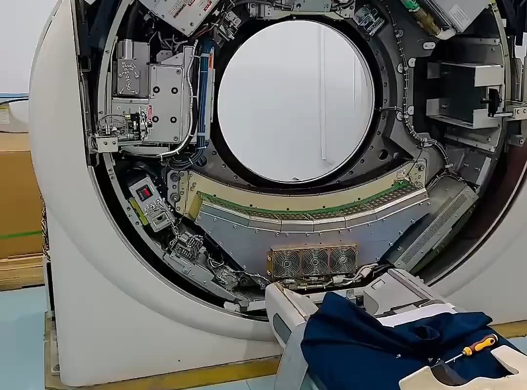
Key Components Inside the CT Scanner
Here’s a breakdown of what you’d find if you could peek inside a CT scan machine:
1. The Gantry: The Heart of the Scanner
The large, donut-shaped structure you see in CT rooms is called the gantry. Inside it, two critical components rotate at high speed around the patient:
- X-ray Tube: Generates the radiation beam that passes through the body.
- Detectors: Measure how much of the X-ray beam is absorbed by tissues. These detectors are directly opposite the tube and capture data thousands of times per second.
Modern CT scanners use slip-ring technology, enabling continuous rotation without tangled wires, which speeds up the scan.
2. The Patient Table
The motorized bed slides the patient into the gantry’s circular opening. Precision motors ensure the table moves incrementally (or remains stationary) to capture images slice by slice. Newer machines can complete whole-body scans in seconds, reducing motion blur.
3. X-ray Generator & Cooling System
High-voltage generators power the X-ray tube, while advanced cooling systems (like oil or air-based) prevent overheating during prolonged use.
4. Detector Arrays
Modern detectors use solid-state materials (e.g., cadmium tungstate or rare-earth ceramics) that convert X-rays into electrical signals. The latest machines feature multi-slice detectors, capturing up to 320 slices per rotation for ultra-high-resolution imaging.
5. Computer System & Software
Raw data from the detectors is sent to a powerhouse computer that reconstructs images using algorithms like FBP (Filtered Back Projection) or iterative reconstruction. These turn thousands of data points into visual slices measured in Hounsfield Units (HU), which differentiate tissues (e.g., water = 0 HU, bone = 1000 HU).
6. Control Room & Shielding
Operators work from a shielded control room, monitoring the scan via computers. Lead-lined walls and glass protect staff from scattered radiation.
How Does a CT Scan Work? Step by Step
- Patient Positioning: The patient lies on the table, which moves into the gantry.
- X-ray Emission: The tube rotates around the body, emitting narrow X-ray beams.
- Data Capture: Detectors measure beam attenuation (how much the X-rays weaken as they pass through tissues).
- Image Reconstruction: The computer assembles this data into cross-sectional slices, which can be stacked to create 3D models.
Fun Fact: A modern CT scanner can complete a full 360-degree rotation in under 0.3 seconds!
Safety Measures Inside the Scanner
- Radiation Shields: The gantry and housing minimize radiation leakage.
- Collimators: These narrow the X-ray beam to target only the area of interest, reducing unnecessary exposure.
- Adaptive Dose Control: Software adjusts radiation doses based on the patient’s size and tissue density.
Why Use a CT Scanner?
- Speed: Critical for emergencies like strokes or internal injuries.
- Detail: Reveals tiny abnormalities missed by X-rays or ultrasounds.
- Versatility: Used in cancer staging, vascular studies, and even virtual colonoscopies.
FAQs About CT Scans
Q: What’s it like to be inside the scanner?
A: The tunnel is open at both ends (unlike an MRI), so claustrophobia is rare. You might hear whirring or clicking as the gantry rotates.
Q: What’s with the IV contrast?
A: Iodine-based contrast agents are often injected to highlight blood vessels or tumors, making them easier to see.
The Future of CT Technology
Advancements continue to push boundaries:
- Photon-Counting CT: Higher resolution with lower radiation doses.
- AI-Driven Analysis: Machine learning helps spot patterns in scans faster.
- Portable & Compact Scanners: Revolutionizing emergency and battlefield medicine.
Conclusion
The CT scan machine is a marvel of engineering, blending physics, computing, and medical expertise to save lives. Next time you see one, you’ll appreciate the intricate dance of X-rays, detectors, and software happening beneath its sleek exterior.
By demystifying how a CT scanner works, we hope to ease anxieties and highlight the importance of this imaging powerhouse. Always consult your doctor to understand if a CT scan is right for your diagnosis.
Meta Description: Discover how a CT scan machine works, its key components, and the science behind its life-saving imaging. Learn how X-rays and computer tech create 3D views of your body!
Target Keywords: CT scan machine, CT scanner components, how CT scans work, CT scan process, CT imaging technology