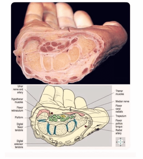
Anatomy of a Human hand compared to that in a textbook.
Meta Title: Human Hand Anatomy Explained: Real-Life vs. Textbook Depictions | Structure & Insights
Meta Description: Discover how the anatomy of a human hand compares to textbook diagrams. Dive into bones, muscles, nerves, and functional complexity in this detailed guide.
Anatomy of the Human Hand: Real-Life Complexity vs. Textbook Simplicity
The human hand is a marvel of biological engineering, enabling precision, strength, and dexterity. While textbooks provide foundational knowledge, real-life hand anatomy reveals nuances often overlooked in diagrams. This article explores the intricate structure of the hand, comparing clinical reality to idealized textbook representations—essential knowledge for students, artists, and medical professionals.
1. The Skeletal Framework: More Than Just 27 Bones
Textbook View:
Textbooks simplify the hand’s skeleton as 27 bones grouped into carpals (8), metacarpals (5), and phalanges (14). Diagrams often depict these as static, perfectly aligned structures.
Real-Life Complexity:
- Variability: Carpal bones like the scaphoid and lunate can vary slightly in shape between individuals.
- Dynamic Alignment: Bones shift during motion (e.g., when making a fist), impacting grip mechanics.
- Clinical Nuance: Fractures in the scaphoid bone are often missed in X-rays due to overlapping shadows—a detail rarely highlighted in textbooks.
2. Muscles and Tendons: Hidden Layers of Control
Textbook View:
Diagrams categorize muscles as extrinsic (forearm-based) or intrinsic (hand-based), with color-coded tendons for clarity.
Real-Life Complexity:
- Interconnectivity: Tendons like the flexor digitorum profundus don’t work in isolation; they sync with lumbricals and interossei for coordinated movement.
- Adaptive Tension: Real hands demonstrate how tendons adjust tension dynamically—think of a pianist’s rapid keystrokes versus a weightlifter’s sustained grip.
- Variations: Some people have “accessory” tendons (e.g., a duplicated palmaris longus), absent in standard textbooks.
3. Nerves and Blood Supply: The Living Highway
Textbook View:
Nerves (median, ulnar, radial) and arteries (palmar arches) are shown as symmetrical, linear pathways.
Real-Life Complexity:
- Branching Patterns: The ulnar nerve’s sensory branches diverge unpredictably in 15% of people—critical for surgeons avoiding nerve damage.
- Blood Flow Dynamics: Textbook arches are idealized; in reality, collateral circulation varies, affecting recovery from injuries.
- Sensory Overlap: Fingertips have overlapping nerve innervation, explaining why localized pain can be hard to pinpoint.
4. Skin and Connective Tissues: Beyond Surface-Level
Textbook View:
Skin is depicted as a uniform layer with simple creases (e.g., palmar flexion lines).
Real-Life Complexity:
- Dermatoglyphics: Fingerprints (friction ridges) are genetically unique and functional, enhancing grip—not just for identification!
- Fascial Layers: Connective tissues like the palmar aponeurosis anchor skin to deeper structures, vital for grasping objects securely.
5. Joints and Movement: A Symphony of Motion
Textbook View:
Joints are labeled as hinge (interphalangeal) or condyloid (metacarpophalangeal), with plain arrows showing motion range.
Real-Life Complexity:
- Compound Movements: Thumb opposition involves the carpometacarpal (CMC) joint rotating, swiveling, and flexing simultaneously—far beyond textbook arrows.
- Ligament Elasticity: Collateral ligaments tighten in flexion but loosen in extension, stabilizing joints without restricting motion.
Why Textbooks Simplify Anatomy
Textbooks prioritize clarity over complexity to help learners grasp basics. However, this can lead to misconceptions:
- Oversimplified Blood Supply: Diagrams omit variations like the “median artery” persistence in 10% of adults.
- Static vs. Dynamic: Diagrams freeze motion, while real anatomy relies on kinetic chains (e.g., wrist position affecting finger strength).
Bridging the Gap: From Diagrams to Reality
To truly understand hand anatomy:
- Dissection & 3D Models: Study cadaveric hands or digital tools like Complete Anatomy™.
- Clinical Imaging: Compare textbook diagrams to MRI/CT scans showing tissue layers.
- Functional Tests: Observe how your own hand moves when typing, gripping, or mimicking injuries.
Conclusion
The anatomy of the human hand is far more intricate than textbook illustrations suggest. While diagrams excel at teaching foundational concepts, real-world variability, dynamic function, and biological adaptability make the hand a masterpiece of evolution. Whether you’re a medical trainee, artist, or curious learner, recognizing this gap enriches your appreciation of human biomechanics.
Keywords: Human hand anatomy, hand bones and muscles, textbook vs real anatomy, hand nerve supply, hand joint mechanics, functional hand anatomy, clinical anatomy variations.
Optimization Tips:
- Link to interactive 3D anatomy platforms.
- Include alt-text for images describing anatomical comparisons.
- Use internal links to related content (e.g., wrist anatomy or nerve injury guides).
- Target long-tail keywords like “how do real hands differ from textbook diagrams?”
By merging academic foundations with real-world insights, this article bridges classroom learning and clinical expertise—perfect for SEO and reader engagement!