Human eye under a microscope.
Unlocking the Hidden World: The Human Eye Under a Microscope
The human eye is a marvel of biological engineering, capable of capturing light, decoding colors, and transmitting intricate visual data to the brain. But what happens when we zoom in beyond what’s visible to the naked eye? Under a microscope, the eye transforms into a landscape of intricate layers, specialized cells, and astonishing structures that reveal just how complex this sensory organ truly is.
In this article, we’ll explore the microscopic anatomy of the human eye, uncover how scientists study it, and explain why these tiny details hold monumental importance for medicine, research, and our understanding of vision.
Why Examine the Eye Under a Microscope?
Studying the eye at a microscopic level helps researchers:
- Diagnose diseases like glaucoma, diabetic retinopathy, and macular degeneration.
- Understand how light-sensitive cells communicate with the brain.
- Develop advanced treatments, including retinal implants and gene therapy.
- Appreciate the eye’s self-healing mechanisms and vulnerabilities.
Preparing the Eye for Microscopy
To observe eye tissues, scientists use a process called histology:
- Fixation: Preserving the tissue (often with formaldehyde) to maintain structure.
- Sectioning: Slicing ultra-thin layers (1–10 micrometers thick) using a microtome.
- Staining: Applying dyes like hematoxylin (highlights nuclei) and eosin (stains proteins) to enhance contrast.
Advanced techniques, such as electron microscopy or immunofluorescence, can pinpoint proteins, nerve connections, and cellular anomalies invisible under standard light microscopes.
Microscopic Anatomy: A Layer-by-Layer Breakdown
1. Cornea: The Transparent Protector
Under the microscope, the cornea reveals five distinct layers:
- Epithelium: Outer skin-like cells that repel dust and germs.
- Bowman’s Layer: A dense mesh of collagen.
- Stroma: Water-rich collagen fibers that refract light (making up 90% of the cornea).
- Descemet’s Membrane: A basement membrane for endothelial cells.
- Endothelium: Pumps excess fluid to prevent corneal swelling.
Microscopic surprise: Corneal stem cells at the edge regenerate the epithelium every 7–10 days!
2. Lens: The Flexible Focuser
The lens is made of tightly packed, transparent cells called lens fibers, filled with crystallin proteins. Unlike most cells, lens fibers lose their nuclei during development to optimize light transmission. Under high magnification, these fibers resemble concentric circles—like layers of an onion.
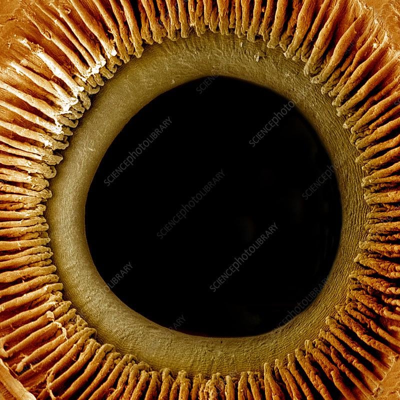
3. Retina: The Photoreceptor Powerhouse
The retina steals the show under a microscope, revealing 10 layers stacked like a neural circuit board:
- Photoreceptors (Rods & Cones): Rods (shaped like cylinders) detect low light; cones (tapered) sense color.
- Bipolar & Ganglion Cells: Relay signals from photoreceptors to the optic nerve.
- Macula Lutea: A yellow-pigmented zone packed with cones for sharp central vision.
- Optic Disc: The “blind spot” where nerve fibers exit—no photoreceptors here!
Electron microscopy even shows mitochondria-packed regions in photoreceptors, fueling their high energy demands.
4. Iris & Pupil: Dynamic Light Regulators
Sections of the iris reveal:
- Melanocytes: Pigment-producing cells that define eye color.
- Sphincter and Dilator Muscles: Contract or expand the pupil in response to light—visible as bands of smooth muscle fibers.
5. Sclera & Choroid: The Eye’s Support System
- Sclera: The “white” outer layer appears as interwoven collagen fibers under magnification, providing structural rigidity.
- Choroid: A vascular layer beneath the retina, packed with blood vessels and melanin (to absorb stray light).
What Microscopy Reveals About Eye Diseases
- Glaucoma: Damaged ganglion cells and a thickened trabecular meshwork (the eye’s drainage system).
- Cataracts: Clumped crystallin proteins clouding the lens.
- Age-Related Macular Degeneration (AMD): Drusen deposits (yellow debris) beneath the retina.
Early detection of these changes can save vision—a key reason microscopy remains vital in ophthalmology.
Beyond the Visible: Advanced Imaging Techniques
- Electron Microscopy: Zooms in 10,000x to reveal synaptic connections between retinal cells.
- Confocal Microscopy: Creates 3D reconstructions of living corneal tissues (used in LASIK pre-screening).
- OCT (Optical Coherence Tomography): Non-invasive imaging that scans retinal layers in real time.
Frequently Asked Questions
Q: Can living eyes be examined under a microscope?
A: Yes! Tools like slit-lamp biomicroscopy allow doctors to inspect the cornea, lens, and retina in real time.
Q: How small are the cells in the eye?
A: Rod and cone cells are just 2–5 micrometers wide—smaller than a human hair (70 μm thick).
Q: Why do some microscope images show eyes in wild colors?
A: Artificial staining or fluorescent dyes highlight specific structures for analysis.
Conclusion: A Universe in Miniature
Peering at the human eye under a microscope is like discovering a hidden cosmos—one where cells collaborate seamlessly to turn photons into perception. As imaging technology evolves, so too does our ability to decode vision’s mysteries, heal damaged eyes, and marvel at nature’s microscopic genius.
Keywords for SEO: Human eye under microscope, microscopic eye anatomy, retinal cells, eye histology, cornea microscopy, ophthalmology imaging, rod and cone cells, electron microscopy eye.
By understanding the microscopic wonders of the eye, we gain not just scientific insight—but a deeper appreciation for the delicate machinery that lets us witness the world.