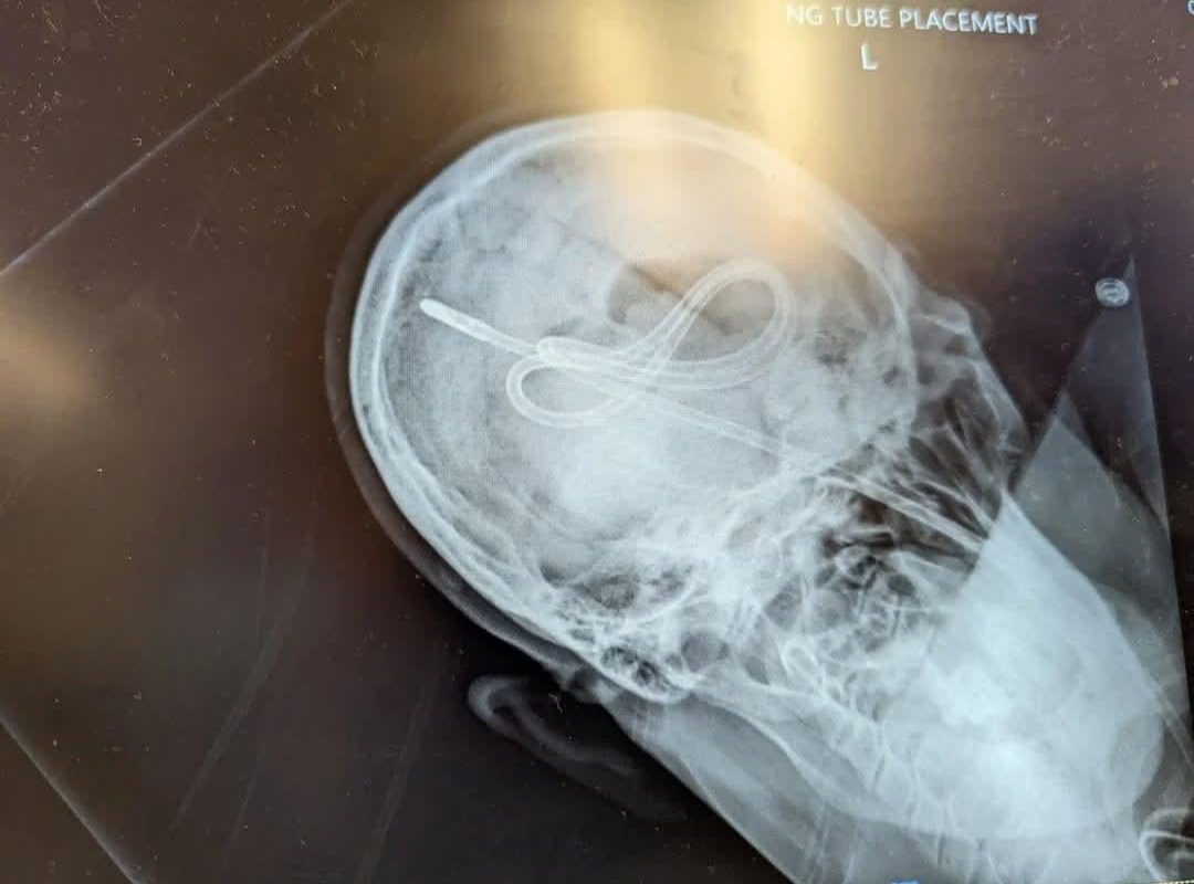
PMCT Images. Nurse advanced NG tube until she heard a pop, then tried an air bolus to ensure placement. Patient did not survive.
Title: PMCT Images: A Critical Insight into Tragic NG Tube Misplacement Fatalities
Meta Description: Explore how PMCT imaging reveals fatal complications of NG tube misinsertion, including a case study where auscultation and air bolus tests failed to prevent a tragic outcome.
Introduction
Nasogastric (NG) tube placement is a routine medical procedure, but errors in confirmation of placement can lead to catastrophic consequences. Post-Mortem Computed Tomography (PMCT), a forensic imaging tool, plays a pivotal role in uncovering these fatal missteps. This article examines a tragic case of NG tube-related death, explores how PMCT imaging exposed the cause, and emphasizes best practices to prevent similar outcomes.
What Is PMCT?
Post-Mortem Computed Tomography (PMCT) is a non-invasive radiographic technique used in forensic investigations to visualize internal injuries, organ damage, or procedural complications after death. Unlike traditional autopsies, PMCT provides 3D imagery of anatomical structures without dissection, making it invaluable for identifying iatrogenic (procedure-related) fatalities.
The Case: NG Tube Misplacement and Fatal Air Bolus
In a tragic incident, a nurse advanced an NG tube into a critically ill patient until hearing an audible “pop.” Believing the sound indicated gastric placement, she administered an air bolus (a common test to confirm tube position via auscultation of whooshing sounds). Shortly after, the patient deteriorated and died.
PMCT imaging later revealed devastating findings:
- Tracheobronchial Perforation: The “pop” signaled unintended tube entry into the trachea or bronchi, causing airway trauma.
- Pneumothorax: The air bolus introduced near the lungs led to a collapsed lung.
- Subcutaneous Emphysema: Air leakage into soft tissues further compromised respiration.
These complications confirmed that auscultation and air bolus tests alone are unreliable for verifying NG tube position.
Why Auscultation & Air Bolus Tests Fail
The tragedy underscores the limitations of traditional NG tube confirmation methods:
- False-Positive Air Bolus Sounds: Air injected into the pleura or esophagus can mimic gastric placement sounds.
- Human Error: Auditory misinterpretations or anatomical variations (e.g., paralyzed vocal cords) increase risk.
- Lethal Complications: Misplaced tubes can perforate organs, induce aspiration, or cause fatal pneumothorax.
PMCT’s Role in Investigating NG Tube Deaths
PMCT imaging provided irrefutable evidence in this case, offering:
- Precise Localization: The tube’s path through the trachea into the pleural cavity was visualized.
- Identification of Secondary Injuries: Air emboli, lung collapse, and soft tissue damage were clearly mapped.
- Forensic Accountability: The images highlighted systemic flaws in procedural protocols.
Without PMCT, the exact mechanism of death might have remained speculative.
Best Practices for Avoiding NG Tube Disasters
To prevent similar fatalities, medical guidelines emphasize:
- pH Testing: Gastric aspirate pH ≤5.5 confirms gastric placement.
- X-Ray Verification: Chest/abdominal X-rays are the gold standard before tube use (per WHO, NPSA).
- Capnography: Detects CO2 to rule out tracheal placement.
- Staff Training: Competency in ultrasound-guided placement and recognition of warning signs (e.g., coughing, hypoxia).
Conclusion: Lessons from PMCT Investigations
PMCT imaging transforms post-mortem investigations by exposing hidden procedural errors. The tragic case of the NG tube misplacement underscores the need to abandon outdated verification methods like air bolus tests. Hospitals must enforce radiological confirmation and prioritize patient safety protocols to avert preventable deaths.
Key Takeaways:
- PMCT provides critical forensic evidence in iatrogenic fatalities.
- NG tube placement requires X-ray or pH testing—never rely on auscultation alone.
- Continuous training and technology adoption save lives.
SEO Keywords:
PMCT images, NG tube complications, nasogastric tube death, post-mortem CT scan, air bolus test failure, tracheal perforation, forensic radiology, NG tube verification, pneumothorax case study.
Internal Links Suggestion:
- “NG Tube Placement Protocols: A Safety Checklist”
- “How Forensic Imaging (PMCT) Revolutionizes Autopsies”
- “Pneumothorax: Causes, Symptoms, and Emergency Management”
By leveraging PMCT insights and adhering to evidence-based guidelines, healthcare providers can turn tragic lessons into life-saving reforms.