The human tongue under very high zoom
The Human Tongue Under Very High Zoom: Revealing a Hidden Microcosmos
When viewed under extreme magnification, the human tongue reveals a surreal, alien-like landscape—one that’s teeming with intricate structures responsible for taste, texture perception, and even speech. Using advanced microscopy, scientists have captured astonishing details of this sensory organ, uncovering a world of papillae, taste buds, and microbial ecosystems invisible to the naked eye. In this article, we’ll zoom in on the tongue’s microscopic architecture, explore its biological marvels, and explain why this “everyday” organ is far more complex than it seems.
The Tongue’s Surface: A Microscopic Jungle
At first glance, the tongue appears smooth but dotted with tiny bumps. Under high magnification, however, it transforms into a dense terrain of papillae—small, specialized projections that give the tongue its rough texture. There are four main types of papillae, each with unique shapes and roles:
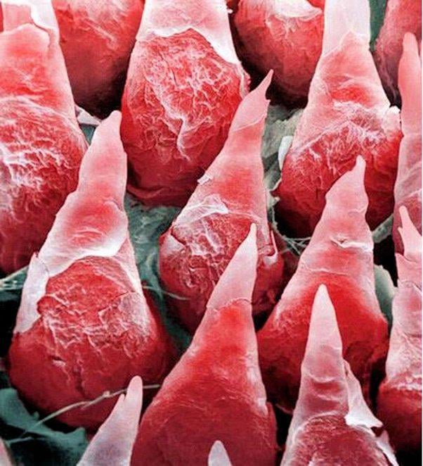
1. Filiform Papillae: The Tiny “Hairs”
- Appearance under zoom: Spike- or thread-like structures covering most of the tongue’s surface.
- Function: These keratinized papillae lack taste buds but act like microscopic brushes, gripping food to aid chewing and swallowing. Under scanning electron microscopy (SEM), they resemble a dense forest of cones or filaments.
2. Fungiform Papillae: The Taste Guardians
- Appearance under zoom: Mushroom-shaped bumps scattered among filiform papillae, often with visible taste buds on their surfaces.
- Function: Each fungiform papilla houses 1–15 taste buds, which detect sweet, salty, sour, bitter, and umami flavors. Under high zoom, these buds look like tiny flower buds embedded in the papillae.
3. Foliate Papillae: The Folded Sensors
- Appearance under zoom: Ridges and grooves along the tongue’s sides, resembling folded fabric.
- Function: These papillae contain hundreds of taste buds clustered in their crevices, making them highly sensitive to flavors.
4. Circumvallate Papillae: The Mighty Moats
- Appearance under zoom: Large, dome-shaped structures encircled by deep trenches (visible only under high magnification).
- Function: Located at the tongue’s rear, these papillae contain thousands of taste buds and salivary glands, flushing flavor molecules toward sensory cells.
Taste Buds Up Close: How Flavor Comes to Life
When magnified, taste buds resemble microscopic onions or barrels. Each bud comprises 50–100 specialized cells:
- Gustatory cells: Detect chemicals in food and send signals to the brain.
- Basal cells: Act as stem cells, regenerating taste buds every 1–2 weeks.
- Supporting cells: Provide structural integrity.
Under SEM, taste buds appear as pores (taste pores) on the papillae’s surface. When food dissolves in saliva, it enters these pores, triggering the cells inside. This process—captured in stunning detail via microscopy—highlights how precision biology translates a bite of pizza into a burst of flavor.
Beyond Taste: Microscopic Ecosystems & Health Clues
Zooming in further reveals more than just taste sensors:
- Microbial Cities: The tongue’s crevices host bacteria, which form biofilms (like dental plaque). Under fluorescence microscopy, these appear as colorful, sticky colonies. A balanced microbiome is crucial for oral health, while imbalances can cause bad breath or disease.
- Diagnostic Potential: Abnormal cell growth (e.g., oral cancer) or infections like oral thrush can be detected early via microscopic tongue analysis.
The Tech Behind the Images: How We See the Unseeable
Capturing the tongue’s microscopic details requires cutting-edge tools:
- Scanning Electron Microscopy (SEM): Uses electron beams to create 3D, high-resolution images at up to 500,000x zoom. SEM reveals papillae and biofilms in extraordinary detail.
- Confocal Microscopy: Laser-based imaging shows living cells and fluorescent-tagged bacteria in real time.
- Atomic Force Microscopy (AFM): Maps surface textures at the nanoscale, even detecting molecular forces on taste buds.
Scientific Insights from a Tiny World
Microscopic studies have led to breakthroughs, such as:
- Identifying super-tasters (people with more fungiform papillae and heightened taste sensitivity).
- Uncovering how spicy foods (via capsaicin) and menthol interact with tongue receptors at a cellular level.
- Mapping taste bud regeneration, offering hope for restoring taste after injury or chemotherapy.
Conclusion: A Testament to Nature’s Precision
The human tongue, when magnified, is a masterpiece of biological engineering—a dynamic sensory hub where taste, touch, and microbiology converge. From the armored filiform papillae to the flavor-decoding taste buds, every microstructure plays a vital role in our daily lives. As imaging technology advances, we’ll continue uncovering secrets of this hidden world, deepening our appreciation for the small wonders within us.
FAQs: The Tongue Under High Zoom
Q: Can you really see taste buds with a microscope?
A: Yes! Light microscopes show taste buds as clusters, while SEM reveals their 3D pore-like structure.
Q: Why does the tongue look prickly under zoom?
A: The “prickles” are filiform papillae, which dominate the tongue’s surface.
Q: How do bacteria appear on the tongue under magnification?
A: They form colorful, clustered biofilms, especially in grooves between papillae.
Q: Does tongue structure vary between people?
A: Yes—genetics affects papillae density, influencing taste sensitivity and texture perception.
Meta Description: Discover the human tongue under extreme magnification! This SEO-optimized article explores taste buds, papillae, and microbial ecosystems revealed via SEM, with FAQs and scientific insights.
Slug: human-tongue-under-high-zoom-microscopic-view