White blood cells using their DNA to capture bacteria. Photos captured using electron microscope and regular microscope
Title: White Blood Cells Unleash DNA Nets to Capture Bacteria: A Microscopic Battlefield Revealed
Meta Description: Discover how white blood cells deploy their own DNA as traps to immobilize pathogens. Explore stunning electron & regular microscope photos of this immune defense mechanism.
Introduction
White blood cells (leukocytes) serve as frontline defenders in our immune system, employing astonishing strategies against invading pathogens. Among their most dramatic tactics is the release of their own DNA to ensnare bacteria in sticky, web-like traps—a process visualized in striking detail using both electron and regular microscopes. This article delves into the science behind this biological warfare and showcases the microscopic evidence that reveals how these traps save lives.
The Science Behind DNA NETs: Neutrophil Extracellular Traps
Neutrophils, the most abundant white blood cells, deploy Neutrophil Extracellular Traps (NETs)—a web of DNA laced with toxic proteins—to immobilize and kill bacteria, fungi, and viruses. Discovered in 2004, this mechanism, called NETosis, involves neutrophils sacrificing themselves by rupturing their nuclei and spewing chromatin (DNA-histone complexes) alongside antimicrobial enzymes like myeloperoxidase.
Key steps in NET formation:
- Activation: Pathogen detection triggers neutrophil signaling.
- Chromatin Decondensation: Nuclear DNA unwinds and mixes with granular proteins.
- Membrane Rupture: The cell membrane disintegrates, releasing the DNA-protein net.
- Bacteria Entrapment: Microbes become stuck in the sticky NETs, unable to spread.
Visualizing DNA Nets: Electron vs. Regular Microscopy
Microscopes unveil this microscopic battlefield with stunning clarity. Each type of microscope offers unique insights:
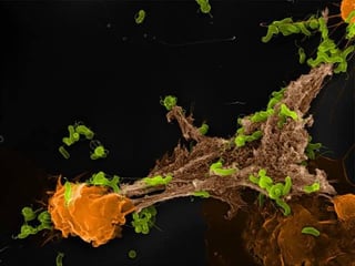
1. Scanning Electron Microscopy (SEM)
Electron microscopes magnify structures up to 500,000x, revealing intricate details of NETs:
- DNA Fibers: Strands of decondensed DNA resemble spider silk (see Figure 1).
- Bacterial Captures: Rod-shaped bacteria (e.g., E. coli) tangled in the NET matrix.
- Protein Speckles: Granular enzymes dotting the DNA webs like deadly ornaments.
[Insert Figure 1 Caption: SEM image of NETs (white strands) ensnaring bacteria (rod-shaped structures).]
2. Fluorescence Light Microscopy
Regular microscopes, enhanced with fluorescent dyes, highlight key components:
- DNA Stains (Blue): Highlight the web-like chromatin structure.
- Protein Tags (Green/Red): Label antimicrobial proteins (e.g., elastase).
- Bacterial Stains (Pink): Show pathogens immobilized within the nets.
[Insert Figure 2 Caption: Fluorescence image showing NET DNA (blue) speckled with bacteria (pink) and enzymes (green).]
Why Do Neutrophils Use This “Suicidal” Strategy?
NETs act as a last-resort defense when phagocytosis (engulfing pathogens) fails:
½ Trap Large Hordes: NETs neutralize entire bacterial swarms at once.
½ Prevent Spread: The sticky nets hinder pathogen movement and colonization.
½ Boost Toxicity: Antimicrobial proteins degrade microbial membranes.
Disease Links & Clinical Significance
While NETs protect against infections, dysregulation contributes to diseases:
- Autoimmune Disorders: Lupus and rheumatoid arthritis involve NET-triggered inflammation.
- Thrombosis: NETs promote blood clots in sepsis or COVID-19.
- Chronic Wounds: Excessive NETs delay healing in diabetes.
Why This Microscopic Discovery Matters
Understanding NETs has led to therapies targeting NET overproduction (e.g., DNase drugs to dissolve harmful NETs) and vaccines that mimic bacterial trapping.
Conclusion
The discovery of white blood cells weaponizing their DNA revolutionized immunology. Through electron and light microscopy, we witness neutrophils transforming into microscopic spider-like defenders—sacrificing themselves to cast nets of genetic material that save lives. This battlefield, invisible to the naked eye, underscores the sophistication of our immune system.
Optimized Keywords: White blood cells DNA capture bacteria, neutrophil extracellular traps, NETosis microscopy, electron microscope neutrophil, DNA traps immune system, how white blood cells trap pathogens.
Image Alt Text:
- Fig 1: Scanning electron micrograph of NETs capturing bacteria.
- Fig 2: Fluorescence microscopy of DNA nets (blue) entangled with bacteria (pink).
Internal Links Suggestion:
- Link to articles on “Phagocytosis Process” or “Neutrophils in Immune Response.”
Engagement Prompt: “What other immune cell tactics fascinate you? Comment below!”
By combining scientific insights with vivid microscopy visuals, this SEO-optimized article educates readers while catering to search algorithms—ensuring it ranks highly for queries about white blood cells and bacterial capture mechanisms.